Table of Contents
Ureteral Duplication, Ureterocele, and Ectopic Ureter
Development of the urinary organs before birth prepares the body to void urine. Sometimes those organs do not develop correctly, perhaps because of heredity or related to an unknown influence. The result is different anatomy that exists at birth—called congenital malformation.
Congenital malformations of the urinary tract include:
These conditions will be defined and discussed here.
Ureteral Duplication
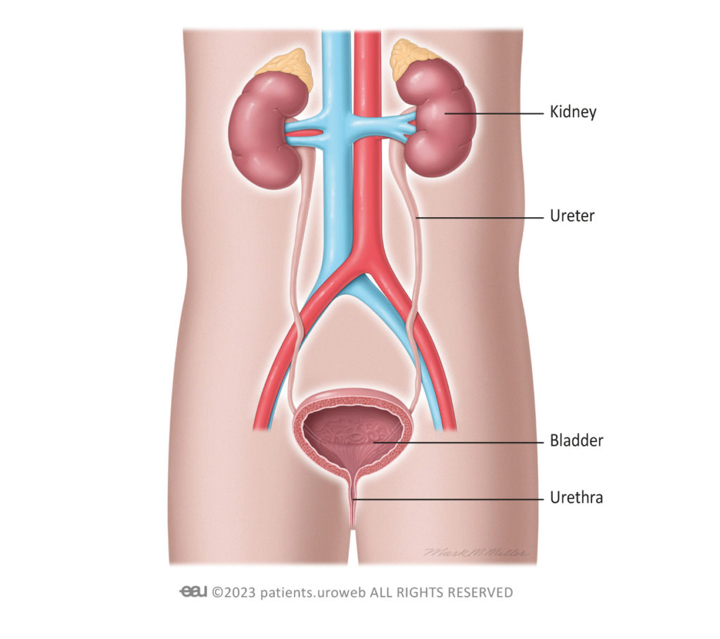
One of the most common malformations of the urinary tract is ureteral duplication. The ureter is the tube that carries urine from a kidney to the bladder. Typically, each kidney has one ureter. In ureteral duplication, however, a kidney has two ureters connecting it to the bladder. This condition is also called a duplicated or duplex collecting system.
Sometimes two ureters may leave the kidney but join into a single tube before arrival at the bladder, it is called incomplete duplication. When the ureters are entirely separate throughout the course to the bladder (Figure 1), it is called complete duplication.
Symptoms and diagnosis of Ureteral Duplication
Despite an extra ureter, a duplicated collecting system can slow the flow of urine, causing urinary obstruction. It is also associated with vesicoureteral reflux (when urine flows backwards from the bladder into the ureter -toward the kidney-), ureterocele and ectopic ureter.
Ureteral duplication is sometimes noticed on routine ultrasound during pregnancy. After birth, symptoms may lead to a diagnosis. However, in the majority of the children with ureteral duplication, no symptoms exist.
Treatment of Ureteral Duplication
Deciding on treatment depends on multiple factors. A conservative approach should always be the first choice whenever possible.
Diagnosis | Definition | Treatment |
Ureteral duplication | A kidney has two ureters connecting it to the bladder instead of one | Monitoring and treatment of symptoms if mild |
Ureterocele
Ureterocele (pronounced “u-ree-tero-seel”) is a pouch-like enlargement of the ureter at the end where it connects to the bladder. This enlargement usually interferes with the flow of urine. Most patients with ureterocele also have a duplicated collecting system. Girls are more likely to be affected.
There are two types of ureterocele (Figures 2 and 3):
- Orthotopic ureterocele: The pouch-like blockage is located completely inside the bladder.
- Ectopic ureterocele: The pouch-like blockage extends into the bladder opening or the urethra. This is the most common form.
Ureterocele usually obstructs the flow of urine into the urethra, the tube that carries urine out of the body. The degree of obstruction varies based on the type of ureterocele and the amount of abnormal tissue development. Ectopic ureterocele is more serious because the bladder opening to the urethra does not work properly.
Symptoms and diagnosis of Ureterocele
Ureterocele symptoms may be noted before or after birth. Ultrasound during pregnancy may show an obstructive ureterocele. If ureterocele is seen on ultrasound before birth, the doctor will repeat the ultrasound to confirm the diagnosis. If ureterocele and a duplicated collecting system are confirmed, the doctor will do further tests to look at kidney function.
Symptoms soon after birth can lead to diagnosis in infants. In girls, the doctor may see a ureterocele in the opening of the urethra at birth. Newborn boys may not be able to pass urine. The kidneys may also be inflamed.
Symptoms that might occur later include bladder inflammation, difficulty urinating, and an urgent need to urinate. Sometimes urine will flow backwards from the bladder into the ureter (toward the kidney) in patients with ureterocele. This condition is called vesicoureteral reflux. Urine flows back into the ureter on the same side as the blockage or into the ureter opposite the blockage.
Treatment of Ureterocele
In most children showing no symptoms (asymptomatic), treatment is not needed and follow-up is recommended. Urinary tract infections are treated with antibiotics, if present. In addition, further tests should be done using x-ray to assess kidney function. If kidney function is good, the ureterocele can be treated using endoscopic decompression. If kidney function is not good, some of the kidney tissue may be removed surgically. This operation is called partial nephrectomy.
Endoscopic decompression of ureterocele
If the upper part of the kidney is working, the ureterocele can be punctured to decompress the pouch-like enlargement blocking the urinary flow. This procedure is called endoscopic decompression. It is performed inside the body through a tube-like surgical instrument called an endoscope. The urologist uses a laser through the endoscope to puncture the ureterocele. This reduces the size of the blockage and helps urine flow. This treatment sometimes causes the urine to flow backwards from the bladder into the ureter (vesicoureteral reflux). This should be treated.
Partial nephrectomy
If the kidney is not working well, surgery may be needed to remove the non-working part.
Treatment
Diagnosis | Definition | Treatment |
Ureterocele | A pouch-like enlargement of the ureter at the end where it connects to the bladder |
|
Ectopic Ureter
An ectopic ureter connects the kidney to a site other than the bladder. This occurs less frequently than ureterocele and is more common in girls. Some patients do not have symptoms, so it can be difficult to tell how often this occurs. Most patients with an ectopic ureter also have complete duplication of the ureter.
The typical locations of ectopic ureters are different in boys and girls:
- In boys, the ectopic ureter never ends below the external sphincter. It often runs from the kidney to the urethra, near the prostate, or near the glands that produce semen.
- In girls, the ureteral opening may be located in the urethra, in the vaginal opening, in the vagina, or in the uterus or fallopian tubes.
Symptoms and diagnosis of Ectopic Ureter
Different symptoms may appear depending on the child’s age and sex:
- In newborns, parents may see pus in the urine (pyuria) or suspect a urinary infection.
- In young girls, parents may see a loss of urine after normal voiding, the presence of urine in the vagina, or another opening for passing urine located near the urethra.
- Preadolescent boys usually have inflammation of the tube that stores and carries sperm (epididymitis) and swollen seminal glands in the pelvis.
If symptoms are noted, the doctor should test if urine flows backwards from the bladder into the ureter (vesicoureteral reflux). In addition, the doctor will test kidney function (DMSA renal kidney scan) to determine whether the area drained by the ectopic ureter is affected. Magnetic resonance urography or high-resolution MRI could be used to study the entire urinary tract.
Treatment of Ectopic Ureter
Ectopic ureter is treated:
- If urine flows backwards from the bladder into the ureter.
- If symptoms occur, such as urinary tract infection, pus in the urine, or kidney inflammation.
- If the upper part of the kidney is not working.
If the kidney is not working well, the doctor may need to perform a partial nephrectomy to remove the nonworking part.
Diagnosis | Definition | Treatment |
Ectopic ureter | A ureter that connects the kidney to a site other than the bladder—for example, to the urethra or the vagina |
|
Overview of treatments congenital malformation of the urinary tract
If symptoms are mild, the doctor might prefer to monitor the condition and treat symptoms such as urinary tract infection and backward flow of urine. Surgical treatments include endoscopic decompression and partial nephrectomy.
Choice of treatment depends on the patient’s condition:
- Patient age
- Overall health
- Kidney function
- Problems with urine flow
- Blockage of urine by a ureterocele
- Parents’ and surgeon’s preferences
Diagnosis | Definition | Treatment |
Ureteral duplication | A kidney has two ureters connecting it to the bladder instead of one | Monitoring and treatment of symptoms if mild |
Ureterocele | A pouch-like enlargement of the ureter at the end where it connects to the bladder |
|
Ectopic ureter | A ureter that connects the kidney to a site other than the bladder—for example, to the urethra or the vagina |
|