What is a ureterocele?
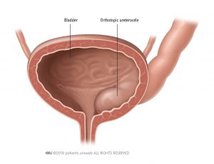
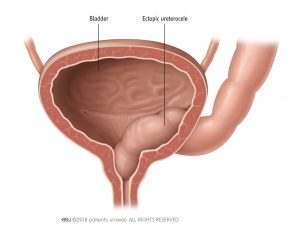
Ureterocele (pronounced: ‘yur-reet-ero-seel’) is a condition where a part of the ureter, the tube that connects the kidney to the bladder, forms a bulge or balloon-like structure inside the bladder. There are 2 types of ureterocele:
- Orthotopic ureterocele, where the bulge is located completely inside the bladder.
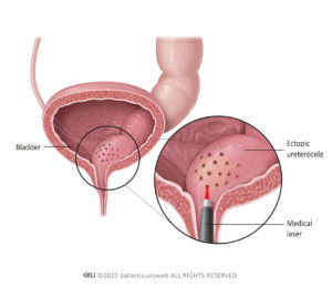
- Ectopic ureterocele, where the bulge extends into the bladder opening or the urethra.
Ureteroceles can cause urine to collect in the balloon-like structure, which can trigger problematic symptoms.
Ureteroceles are quite rare, affecting around one in every 4,000 children. It’s more commonly seen in girls than in boys, with girls being 4 to 7 times more likely to have the condition. About 80% of ureteroceles in children are linked to duplex kidneys, especially those affecting the upper part of the ureter (ureters are the tubes that run down from each kidney to the bladder).
While ureteroceles are not often detected before birth, the condition can cause an enlargement (swelling) of your baby’s urinary tract, which can be seen on an ultrasound scan. This swelling of the urinary tract could be due to a blockage in the flow of urine or be a sign of other urinary tract conditions.
Your medical team will keep a watchful eye on any swelling of your unborn baby’s urinary tract during your pregnancy. This way, if they detect potential problems, they will be on hand to provide specialist healthcare support to ensure your baby receives the best possible care and treatment.
What are the symptoms of a ureterocele?
Usually smaller and non-obstructed ureteroceles don’t cause any symptoms. Not all children with ureterocele experience symptoms and in some cases, the condition is only found when a child is undergoing medical scans or urinary tests later in life. The symptoms of a ureterocele can vary, but they often include:
When a ureterocele creates a bulge where urine can collect, it can result in stagnant or stale urine which can be a perfect place for bacteria to grow. This can lead to a UTI. The main symptom young children with a UTI experience is a high fever. Young infants and toddlers may not show specific UTI symptoms such as pain when weeing, and instead may be irritable or have trouble feeding. Children over the age of 6 years may need to wee a lot, have cloudy or foul-smelling urine, or abdominal discomfort. They are more likely to be able to describe their symptoms.
When a ureterocele blocks the normal flow of urine, it can lead to a backup of urine in the kidney, causing irritation and inflammation. This makes the bladder contract (squeeze) more often. That’s why children with a ureterocele might feel like they have to wee often and urgently. They may try to empty their bladder more often in an attempt to relieve the pressure and discomfort they are feeling.
Haematuria (pronounced ‘hee-mat-yur-ee-ya’) is the medical term for blood in the urine. This symptom is extremely rare in childhood.
In children with a ureterocele, the bulge blocks the normal flow of urine through the ureter (ureters are the tubes that run down from each kidney to the bladder), causing urine to get stuck in the ureter or kidney. This can cause swelling, inflammation (irritation), and bleeding. The increased pressure in the urinary system can make the blood vessels break, which can be another cause of the blood in the urine.
What treatments are available for ureteroceles?
The treatment of a ureterocele depends on how severe your child’s symptoms are and how it affects their kidneys. Regular follow-up appointments with a specialist medical professional are important to manage and monitor the condition.
Non-surgical treatments
In mild cases where the symptoms of a ureterocele are not causing complications, close monitoring by a doctor with regular check-ups may be recommended. This is called a ‘wait and see’ approach. However, if ureterocele is causing your child to keep getting UTIs or suffer kidney-related issues, appropriate treatment options will be discussed with you.
If your child has regular UTIs, your doctor may prescribe antibiotics. These medicines treat the current infection and prevent future ones. Antibiotics kill the bacteria causing the infection and help to make your child feel better.
Surgical Treatments
Your child’s doctor may recommend surgery to treat the ureterocele. The treatment options offered to you will depend on your child’s individual circumstances and where in Europe you live, as healthcare practices differ between countries. Your doctor will discuss all treatment options available, the potential outcomes and any risks with you. Any recommendation they make will be based on careful assessment of your child’s individual medical circumstances.
In the majority of cases, surgery is carried out under general anaesthesia. This is where your child is given medication to send them to sleep. Once they are asleep, they do not feel anything, nor will they remember the operation.
After the procedure, your child will be taken to the recovery area and closely monitored. Your healthcare team will provide instructions on wound care, pain management and any potential dietary or activity restrictions. It’s important to follow these instructions so that your child has a good recovery.
This operation involves having a thin tube with a camera on the end, called an endoscope, passed through the urethra (wee tube) to reach the ureterocele.
The doctor uses the endoscope to see the ureterocele and they can also use it to make a small cut in the ureterocele with a medical laser. The openings created with the laser allow the trapped urine to flow correctly into the bladder, relieving the blockage and preventing further urine from building up.
Endoscopic decompression procedures involve a risk of developing a condition called vesicoureteral reflux (pronounced ‘vessy-co-yur-eet-er-ral ree-flux’), where urine flows backwards from the bladder and back up into the ureters or kidneys. This can happen if a puncture or cut is made in the urinary system during the procedure. If reflux becomes a problem, additional surgery may be necessary.
A less likely scenario, if there is a puncture, is that urine may flow back through the puncture site. Similarly, if there is a cut during the procedure (which happens 40% of the time) urine may flow backward through the cut.

This surgery is carried out if a ureterocele causes severe urinary blockages or frequent UTIs.
The surgeon makes a small cut in the lower abdomen to reach the bladder, then carefully disconnects the affected ureter from the bladder and repositions it to help improve the flow of urine from the kidneys to the bladder and prevent any blockages or reflux of urine.
During the surgery, the surgeon will also remove the ureterocele.
Complete primary reconstruction is a major surgery which involves removing the ureterocele while reconstructing the ureter and the bladder to ensure urine can flow correctly. This procedure is typically recommended for very large and ectopic ureterocele which are causing problems with bladder emptying, or if other treatment methods have been unsuccessful.
The procedure involves a surgeon making a small cut in the lower abdomen and pelvic area to access the ureter and bladder. The ureterocele is then removed and the ureter and bladder are reconstructed to help improve urine flow and prevent future blockages or reflux.
This surgery is used when a kidney (or a part of it) is causing problems such as high blood pressure, severe UTIs, kidney stones, poor functioning, or if there is an issue that is too large to treat with other methods. The surgery involves removing the kidney, the entire ureter connecting it to the bladder, and a small piece of bladder where the ureter and bladder connect, including the ureterocele.
This procedure can be done in a minimally-invasive way such as with laparoscopy/robotic surgery, where small cuts are made and surgical instruments are threaded through, rather than with open surgery (which would involve a large cut being made in the abdomen).


